



# Unlocking the Secrets of Human Tissue Aging Through Computational Histology: A New Framework for Mapping Structural Decline Across the Body
## Introduction
Aging is often perceived as a gradual, uniform process, but recent research reveals that tissue decline follows highly nonlinear trajectories, varying dramatically between organ systems and even within the same individual. Understanding when and how tissues undergo structural remodeling during aging has profound implications for predicting disease risk, timing interventions, and designing personalized geroprotective therapies. Historically, aging research has relied heavily on molecular clocks—tools that estimate biological age from DNA methylation or gene expression patterns. However, these approaches have significant limitations, particularly their assumption of linear change, which fails to capture the complex, nonlinear nature of tissue deterioration.
A team of researchers has now developed a novel computational framework that tackles this challenge by leveraging the rich architectural information encoded in standard histopathology images. By analyzing high-resolution tissue slides from a large cohort of healthy individuals spanning a wide age range, this approach maps out precisely when different organs undergo their most dramatic structural transformations during a lifetime—and reveals that these transformations cluster into distinct waves that vary by tissue type.
## The Computational Framework: Mapping Structural Aging from Histology
The foundation of this approach rests on a simple but powerful premise: the microscopic architecture of tissues—their cellular arrangements, stromal composition, and structural integrity—directly reflects their functional capacity, and changes in this architecture during aging carry critical biological signals. The framework extracts these signals from routine hematoxylin and eosin (H&E) stained slides, the most widely used staining technique in pathology.
### Data and Tissue Scope
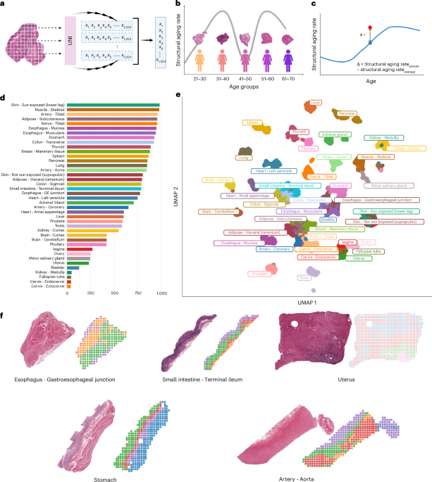
The study drew on post-mortem tissue biopsies from nearly a thousand healthy individuals, ranging in age from their early twenties to their seventies, encompassing samples from forty distinct tissue types across the human body. This generated over twenty-five thousand whole-slide scans, from which more than thirty million individual tissue patches were extracted at high resolution. Histological annotations from the original tissue archive confirmed that age-related changes such as increased atrophy, fibrosis, cyst formation, and atherosclerotic deposits were already present in these curated samples, providing confidence in the quality and relevance of the dataset.
### The Three-Step Analytical Pipeline
The analytical workflow proceeds through three sequential stages, each building upon the previous one to move from raw images to actionable biological insights.
**Step One: Feature Extraction.** Each whole-slide image is first processed to remove background noise, then subdivided into small patches of a standardized size. Every patch is passed through a sophisticated vision model—a type of artificial intelligence architecture known as a vision transformer—that has been pretrained on over one hundred thousand pathology slides. This model converts each patch into a detailed numerical representation, or embedding, capturing over a thousand dimensions of morphological information. By averaging these patch-level representations, the framework generates a single, comprehensive description of each tissue sample’s microscopic structure. Remarkably, these descriptions are powerful enough to automatically distinguish between different tissue types and their substructures without any explicit labeling, demonstrating that the model has learned to capture biologically meaningful architectural features.
**Step Two: Constructing Structural Aging Trajectories.** With slide-level representations in hand for each sample, the next question is temporal: when does a given tissue undergo its most rapid structural changes? The framework addresses this by dividing the age range into overlapping ten-year windows and comparing morphological profiles between adjacent windows. By sliding this window one year at a time, the method constructs a smooth, continuous trajectory of structural aging rates across the lifespan. Periods of pronounced acceleration in structural change are identified as accelerated structural aging (ASA) windows—time points when a tissue is undergoing particularly intense remodeling.
**Step Three: Individual-Level Deviation Analysis.** Once population-level trajectories are established, the framework turns to the individual. For each tissue sample from a specific person, it calculates how much that individual’s structural profile deviates from the expected trajectory for their age. These personalized deviation scores reveal which individuals are aging structurally faster or slower than their peers, opening the door to studying what drives these differences at the clinical, lifestyle, and genetic levels.
## Nonlinear Structural Aging in the Ovary
The ovary presents an ideal test case for this framework because its functional decline is well characterized and distinctly nonlinear. Female fertility begins a gradual decline in the early thirties, accelerates sharply as follicular reserves deplete through the late thirties, and culminates in menopause around fifty to fifty-five years of age. Traditional molecular clocks trained on chronological age assume a linear progression and therefore cannot capture these dramatic nonlinear transitions.
When applied to ovarian tissue, the framework produced a striking bimodal trajectory with two distinct peaks of accelerated structural aging. The first peak occurs between ages thirty-five and forty, aligning precisely with the period of fertility decline. The second peak falls between ages fifty-five and sixty, corresponding to the menopausal transition and the postmenopausal involution of ovarian tissue. These trajectories emerged entirely without any supervision from chronological age—the model simply learned the patterns from the morphological features alone.
Crucially, when the same analysis was applied to gene expression and DNA methylation profiles from the exact same tissue samples, neither molecular data type recapitulated the bimodal pattern. Expression profiles could distinguish young from old ovaries but missed the two-phase structure, while methylation profiles failed to separate young from old samples altogether. This highlights a fundamental advantage of imaging-based approaches: they capture structural information—architectural reorganization, cell loss, fibrosis—that transcriptomic and epigenomic snapshots alone may not reflect.
Further investigation using a vision-language pathology model confirmed that both accelerated periods featured increased fibrosis and tissue atrophy, with a particularly dramatic loss of egg-containing follicular structures during the second period. Pathway analysis of matched molecular profiles revealed that the first period coincided with heightened inflammatory signaling, suggesting a pro-inflammatory microenvironment driving the loss of ovarian reserve, while the second period was marked by the downregulation of growth factor pathways and cell cycle activity, consistent with the cessation of reproductive function after menopause.
## Structural Aging Patterns Across the Human Body
Encouraged by the ovary results, researchers extended the framework to all forty tissue types with sufficient sample sizes, revealing that structural aging follows three fundamentally different temporal programs across organ systems.
### Early Aging Tissues
Some organs undergo their most dramatic structural remodeling during young adulthood, with peak aging rates in the thirties. The vascular system exemplifies this pattern: both tibial and coronary arteries show their steepest structural changes during this decade. This early vascular aging aligns with findings from proteomic studies and is corroborated by pathology annotations showing that atherosclerotic plaque formation accelerates most sharply between the twenties and thirties before plateauing in later decades. Interestingly, the tibial nerve also displays early structural aging—a finding that challenges assumptions about neural resilience and warrants further investigation.
### Late Aging Tissues
Other tissues remain structurally stable for decades before undergoing major transformation in later life. The uterus and vagina follow this late-aging pattern, with their highest remodeling rates concentrated in the early to mid-fifties. This timing corresponds directly to menopause, during which the uterus undergoes dramatic atrophy, endometrial thinning, and myometrial restructuring following the withdrawal of estrogen. The vagina and uterus appear to coordinate their structural aging with the ovary’s late ASA period, suggesting that reproductive aging is a system-wide phenomenon driven by shared hormonal regulation.
### Biphasic Aging: The Predominant Pattern
The most common pattern observed across tissues was biphasic aging—two distinct waves of accelerated structural change separated by a period of relative stability. Nine out of fourteen analyzed tissues, including the esophagus, stomach, colon, small intestine, salivary gland, prostate, testis, and tibial artery, all exhibited this biphasic signature. The first wave typically peaked in the thirties and the second around the fifties, though the exact timing and intensity varied by tissue.
A critical finding was that post-mortem artifacts—such as differences in tissue preservation quality, RNA integrity, and ischemic time—explained negligible variance in these structural aging trajectories, accounting for less than ten percent of the variation in nearly all tissues examined. This provides strong evidence that the observed patterns reflect genuine biological aging processes rather than technical confounders.
The trajectories also proved robust to the choice of analytical model. When alternative pretrained pathology models were used in place of the primary encoder, the same fundamental patterns—early aging for vascular tissues, late aging for reproductive organs, and biphasic aging for digestive and male reproductive tissues—were reproduced across most tissue types.
## Molecular Programs Underlying Accelerated Structural Aging
Having mapped when structural aging occurs in different tissues, the researchers next asked what molecular processes drive these periods of accelerated remodeling. By comparing gene expression profiles from each ASA window to the preceding stable period, a coherent molecular picture emerged.
### A Shared Inflammatory and Metabolic Decline Signature
Across virtually all tissues, accelerated structural aging periods were characterized by a common molecular program. Inflammatory pathways—including TNF signaling, interferon responses, and complement activation—were broadly upregulated. Simultaneously, pathways responsible for energy production (oxidative phosphorylation), cellular proliferation (cell cycle and growth factor targets), and quality control (protein folding, DNA repair, and metabolic regulation) were suppressed. This combination—heightened inflammation alongside declining regenerative and maintenance capacity—appears to be the universal molecular signature of structural aging acceleration.
### Tissue-Specific Vulnerabilities
Within this shared program, individual organ systems displayed distinct molecular weaknesses. In arteries, the peroxisome pathway—a critical process for fatty acid oxidation, protective lipid synthesis, and detoxification of reactive oxygen species—was uniquely downregulated, with the decline restricted to the aorta and tibial artery and absent from all other tissues. This artery-specific loss of peroxisomal function may compound early cardiovascular aging by promoting lipid accumulation and oxidative damage in vessel walls.
The testis exhibited the most dramatic molecular response of any tissue, with widespread upregulation of inflammatory and immune pathways alongside the strongest suppression of spermatogenesis. This pattern is consistent with immune-mediated destruction of germ cells following breakdown of the blood-testis barrier, suggesting that male reproductive aging may involve an inflammatory catastrophe rather than a simple hormonal decline.
### A Temporal Shift from Hormonal Signaling to Damage Response
Comparing the first and second periods of accelerated aging revealed a systematic molecular transition. Hormone-responsive pathways—including estrogen and androgen signaling—declined between ASA-1 and ASA-2 in the vast majority of tissues examined, indicating a broad loss of hormonal responsiveness as tissues progress through their structural aging timeline. Strikingly, this decline was not limited to reproductive organs; gastrointestinal tissues, which express estrogen receptors throughout their epithelial linings, showed the steepest drops in hormone pathway activity.
Meanwhile, damage-response pathways moved in the opposite direction. The unfolded protein response, reflecting increasing accumulation of misfolded proteins, activated in most tissues during the second ASA period. DNA repair pathways, responding to rising genotoxic stress, and reactive oxygen species pathways, signaling escalating oxidative damage, were similarly upregulated. This temporal shift suggests that the body’s first wave of structural aging is driven primarily by inflammatory and hormonal changes, while the second wave is dominated by the cumulative burden of molecular damage that outpaces repair mechanisms.
## Coordinated Aging Across Organs Within Individuals
Perhaps the most intriguing finding concerns how aging is coordinated across different tissues within the same person. By computing structural aging deviation scores for each tissue of each individual and correlating them across tissue pairs, the researchers discovered clear patterns of synchronized deterioration.
Tissues within the same organ system showed strong positive correlations: individuals who aged faster in their colon also tended to age faster in their esophagus and stomach. The same held for vascular tissues—accelerated aging in one artery was mirrored in others—and for female reproductive organs, where uterine and vaginal structural aging moved in tight coordination. Brain regions also showed coordinated aging, and cardiac aging correlated broadly across all tissue types examined.
Beyond these within-system correlations, the researchers identified a surprising cross-system axis linking the digestive and reproductive tissues. Individuals showing accelerated aging in the colon and esophagus also exhibited faster structural aging in the prostate. This coordination aligns with the shared biphasic aging pattern of these tissues and is supported by the molecular finding that gastrointestinal tissues show particularly strong enrichment of hormone-responsive pathways during their first ASA period—even stronger than reproductive tissues themselves. These results suggest that declining hormonal signaling may serve as a systemic coordinator of structural aging, linking deterioration across anatomically distant organs through a shared regulatory axis.
Additional cross-system coordination patterns were observed involving neurovascular coupling—where tibial nerve aging correlated with arterial aging—and sex-specific neuroendocrine coordination, with pituitary-brain correlations being stronger in male individuals than in females. The biological mechanisms driving these axes remain an open area for future investigation.
## Clinical and Genetic Drivers of Structural Aging
### Clinical Associations
To determine whether accelerated structural aging has measurable clinical consequences, the researchers tested associations between individual-level structural aging deviation scores and medical records. The results were striking: accelerated structural aging showed strong positive associations with virtually every canonical aging pathology examined. Atrophy was broadly associated across tissues, particularly in the gastrointestinal and reproductive systems. Fibrosis, vascular calcification, and hyalinization were all linked to elevated structural aging scores. Systemic diseases including autoimmune conditions and neurological disorders showed positive associations across multiple tissues, confirming that individuals with these diagnoses exhibit globally accelerated structural aging.
### Genetic Determinants
A genome-wide association study linking germline genetic variants to structural aging scores identified one hundred twenty-three genes with significant associations. The pathways enriched among these genetic hits directly mirrored the molecular programs discovered through the trajectory analysis: variants that accelerate structural aging were concentrated in oxidative phosphorylation pathways, aligning with the universal decline in energy production during ASA periods. Conversely, protective variants were enriched in immune signaling pathways, particularly those involving interleukin-17 and interferon signaling—suggesting that the genetic capacity to modulate the inflammatory response, the most consistently upregulated program during structural aging acceleration, confers resilience against it.
The most compelling individual genetic finding involved the SIRT6 gene. Variants in SIRT6—a NAD-dependent deacetylase and master regulator of DNA repair, telomere maintenance, and metabolic homeostasis—were specifically associated with accelerated structural aging in vascular tissues. This vascular specificity mirrors the molecular finding that arteries alone lose peroxisomal function during ASA periods, a vulnerability rooted in the same NAD-dependent metabolic processes that SIRT6 governs. Animal studies have shown that SIRT6 knockout produces early atherosclerosis, and these human findings provide a striking translational link between a known longevity regulator and organ-specific structural aging. This result underscores a broader principle: aging pathways do not act uniformly across the body but have tissue-selective effects, meaning that interventions targeting specific organs may need to be tailored accordingly.
## Frequently Asked Questions
### What is the PathStAR framework and what does it do?
PathStAR is a computational method that analyzes high-resolution histopathology images to quantify when and how tissue structure changes during aging. It extracts morphological features from tissue biopsies, constructs population-level structural aging trajectories, and calculates individual-level deviation scores to identify people whose tissues are aging faster or slower than expected.
### How does this approach differ from traditional molecular clocks?
Traditional molecular clocks estimate biological age from DNA methylation or gene expression data and generally assume linear aging. PathStAR is fundamentally different because it operates on histological images—capturing tissue architecture directly—and does not assume linearity. It can detect nonlinear, phase-based patterns of structural remodeling that molecular approaches miss entirely.
### Why is the ovary a particularly interesting case study?
The ovary has a well-established pattern of nonlinear functional decline: gradual fertility loss through the thirties, rapid depletion through the late thirties, and menopause in the early fifties. Because this pattern is so clearly defined, it serves as an excellent validation test for the framework, demonstrating that histology-based analysis can capture established nonlinear biological transitions without being explicitly trained on chronological age.
### What are the three patterns of structural aging observed across tissues?
The three patterns are: (1) early aging, where structural changes peak in the thirties and then decline—seen in vascular tissues; (2) late aging, where tissues remain stable until major remodeling occurs in the fifties or sixties—seen in the uterus and vagina; and (3) biphasic aging, where two distinct waves of acceleration occur, typically in the thirties and fifties, separated by a stable period—this was the most common pattern, observed in nine of fourteen analyzed tissues.
### How were post-mortem artifacts controlled for?
The researchers systematically tested whether technical factors such as tissue preservation quality (RNA integrity number), autolysis, and ischemic time could explain the observed structural aging patterns. These artifacts accounted for less than ten percent of the variance in structural aging rates across nearly all tissues, providing strong evidence that the results reflect genuine biological aging.
### What did the molecular analysis reveal about the difference between the first and second periods of accelerated aging?
The first period of accelerated structural aging is characterized primarily by inflammatory activation and hormonal signaling changes. The second period is defined by a shift toward damage-response pathways, including increased protein stress, DNA repair activity, and oxidative damage—suggesting that cumulative molecular damage becomes the dominant driver of structural aging in later life.
### What is the significance of the SIRT6 finding?
SIRT6 variants were found to be specifically associated with accelerated structural aging in arteries, not in other tissues. This vascular specificity connects a known longevity gene to a tissue-selective aging vulnerability, demonstrating that genetic factors can influence aging in an organ-specific manner rather than uniformly across the body.
### Can lifestyle factors explain differences in structural aging?
In this study, lifestyle variables did not reach statistical significance, likely due to limited sample sizes for certain exposures. This does not mean lifestyle is unimportant for structural aging, but rather that the study was not powered to detect these associations. Larger studies would be needed to evaluate environmental and behavioral contributors.
### Does this framework work with different AI models?
Yes. When alternative pretrained pathology models were tested, the same fundamental aging patterns—early aging for vascular tissues, late aging for reproductive organs, and biphasic aging for digestive tissues—were reproduced, indicating that the findings are robust and not artifacts of a specific model choice.
## Conclusion
The development of computational frameworks capable of mapping structural aging from routine histopathology images represents a significant advance in our understanding of how the human body deteriorates over time. By revealing that tissue remodeling follows nonlinear, organ-specific trajectories—often organized into two or three distinct waves—this work challenges the longstanding assumption of uniform, linear aging and opens new avenues for precision interventions.
The identification of a shared molecular signature of accelerated structural aging, combined with tissue-specific vulnerabilities and cross-organ coordination patterns, provides a rich foundation for understanding why certain organs age faster than others and why some individuals are more resilient than their peers. The convergence of genetic findings with molecular trajectory data reinforces the idea that aging pathways have tissue-selective effects, meaning that effective anti-aging strategies may need to be organ-targeted rather than applied broadly.
Looking ahead, this framework could be applied to clinical biopsy samples to assess individual biological aging status, predict disease risk before symptoms appear, and monitor the effectiveness of interventions designed to slow or reverse specific aspects of tissue deterioration. As larger histopathology datasets become available and imaging technologies continue to improve, the ability to extract actionable aging intelligence from standard tissue slides will only grow, bringing us closer to a future where structural aging can be measured, tracked, and potentially modulated throughout the human lifespan.
Thank you for reading
