



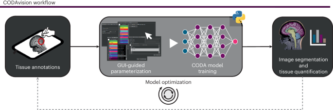
**Harnessing Artificial Intelligence in Digital Pathology: Current Applications and Future Directions**
Digital pathology, the digitization of glass slides into high-resolution images enabling computational analysis, is undergoing a profound transformation through the integration of artificial intelligence (AI) and deep learning. This synergy is paving the way for more accurate, efficient, and personalized diagnostics, addressing long-standing challenges in pathology such as subjectivity, time-consumption, and the increasing complexity of data interpretation. This article synthesizes key insights from recent advances in AI-driven medical image analysis, particularly within the realm of digital pathology.
**The Evolution and Impact of Deep Learning in Pathology**
Deep learning, a subset of machine learning involving neural networks with multiple layers, has demonstrated remarkable success in medical image analysis, including pathology. Its ability to automatically learn hierarchical features from large datasets makes it exceptionally suited for tasks such as image classification, segmentation, and object detection. In pathology, this translates to the potential to augment and even surpass human performance in certain diagnostic tasks, leading to the concept of a “third revolution in pathology” driven by AI. Early foundational work has shown deep learning’s capability in cancer detection, grade prediction, and prognostication across various cancer types, including prostate and pancreatic cancers. The integration of these AI tools into clinical workflows promises to enhance diagnostic precision, support personalized treatment planning, and ultimately improve patient outcomes.
**Key Applications and Technological Frontiers**
One of the most significant applications is in cancer diagnosis and risk stratification. AI algorithms can analyze whole-slide images (WSIs) to identify subtle morphological patterns indicative of malignancy, pre-cancerous lesions, or specific genetic alterations, often with greater speed and consistency than the human eye. For instance, AI-assisted gland analysis in prostatectomy samples has enabled more precise non-destructive 3D pathology and risk stratification. Furthermore, the field is rapidly evolving with the incorporation of multimodal data. Deep learning models are being developed to integrate histology images with spatial transcriptomics data, creating a more comprehensive molecular and cellular understanding of the tumor microenvironment. This integration allows for the elucidation of complex spatial relationships between cancer cells, immune cells, and stroma, which is critical for understanding tumor behavior and response to therapy.
**Emerging Tools and Frameworks**
A burgeoning ecosystem of tools and frameworks is democratizing access to these advanced techniques. Open-source platforms like **QuPath** have become standard for digital pathology image analysis, providing a robust foundation for cell segmentation, annotation, and machine learning integration. Similarly, **OpenSlide** offers a vendor-neutral foundation for viewing and analyzing whole-slide images, ensuring interoperability across different imaging systems. On the more specialized end, frameworks like **Cellpose** serve as a generalist algorithm for cellular segmentation in various biomedical imaging contexts, while **Stardist** leverages deep learning for instance segmentation of cells. The development of user-friendly environments, such as **DeepImageJ**, which allows researchers to run deep learning models directly within the ImageJ interface, further lowers the barrier to entry for implementing these advanced analytical methods.
**Challenges and Future Directions**
Despite the immense promise, several challenges remain. The creation of high-quality, large-scale, and well-annotated datasets is a primary hurdle. The “black box” nature of some deep learning models also raises concerns regarding interpretability and trust in clinical settings. Ensuring algorithmic fairness, robustness, and generalizability across diverse patient populations and imaging equipment is paramount. Future research is actively addressing these issues through the development of more data-efficient learning methods, weakly supervised learning paradigms, and the creation of standardized reporting guidelines for AI in pathology. The convergence of high-resolution 3D imaging technologies, such as serial sectioning and advanced clearing methods, with AI-driven analysis is expected to unlock unprecedented insights into tissue architecture and disease progression.
In conclusion, artificial intelligence is not merely an adjunct in digital pathology but a transformative force. By automating complex image analysis tasks and uncovering hidden patterns within rich multimodal data, AI is helping to realize the vision of precision pathology. As these technologies mature and integrate seamlessly into clinical practice, they hold the potential to revolutionize cancer diagnosis, improve patient stratification for therapy, and accelerate fundamental biomedical discovery.
—
**Original Source Article:** The information and references in this article are derived from the comprehensive review and citation list provided in the original publication: *A multifaceted review on the current state and future of deep learning in medical image analysis*, which compiled an extensive collection of foundational and recent studies (Refs 1-69) detailing the advancements in deep learning for medical imaging, with a specific focus on pathological applications.
