



# How Self-Supervised AI Is Transforming Our View of Living Cells
## A New Way to Teach Computers About Biology
Scientists have long sought ways to teach computers how to “see” and understand living cells. One powerful approach, called Cell Painting, uses fluorescent dyes to light up different parts of a cell—its nucleus, mitochondria, skeleton-like structures, and more. By capturing detailed images of these stained cells, researchers can create rich profiles that reveal how a cell changes in response to thousands of different drugs or genetic modifications.
But analyzing millions of these images by hand is impossible. This is where artificial intelligence steps in. Recently, a new generation of AI models trained through a technique called self-supervised learning (SSL) has shown remarkable promise in extracting meaningful biological information from these images—without needing labels for every single sample.
## Building a Massive Library of Cell Images
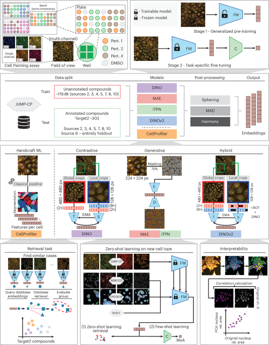
To train these AI models, researchers compiled the largest publicly available collection of Cell Painting images ever assembled. This library contains roughly 5.8 million detailed images of human bone cancer cells, each captured across five different fluorescent channels. The cells were exposed to about 136,000 different chemical and genetic treatments, generated across multiple independent laboratories worldwide.
These images were captured at high resolution, revealing intricate details of cellular architecture. The diversity of treatments, laboratories, and experimental conditions makes this library an extraordinarily rich training ground for AI models.
To further test and validate their models, researchers also worked with three additional datasets. One contained over 2,400 carefully annotated drug compounds tested on two different human cell types across four imaging centers. Another dataset focused on breast cancer cells treated with various chemicals, with each treatment labeled by its known mechanism of action. A fourth dataset featured gene-silencing experiments across four distinct human cell types, providing a valuable benchmark for evaluating how well AI models generalize to new biological contexts.
## Teaching AI to Learn Without Labels
The key innovation behind these models lies in self-supervised learning. Instead of requiring scientists to manually label every image with its biological meaning, SSL allows the AI to learn by observing patterns within the data itself.
Several SSL strategies were explored. One approach, inspired by contrastive learning, teaches the AI to recognize that different cropped views of the same cell image—despite appearing slightly different due to random transformations—belong together, while views of different cells should be kept apart. Another strategy, called masked learning, hides portions of an image and trains the AI to reconstruct what is missing, much like a digital puzzle solver.
A particularly advanced technique combines both contrastive and masked learning into a single framework, using a “student” network that learns from a “teacher” network. The teacher is gradually updated to reflect what the student has learned, creating a feedback loop that produces increasingly robust understanding.
All of these models are built on a backbone architecture known as a Vision Transformer, which breaks each image into small square patches and processes them together, allowing the AI to capture both fine-grained local details and broader global patterns.
## Preprocessing: Making Images AI-Ready
Before feeding images into these models, careful preprocessing is essential. Each image undergoes intensity adjustments to ensure consistent brightness and contrast across different imaging systems. The images are also cropped into smaller, non-overlapping patches using a regular grid pattern, with special care taken to avoid empty regions that contain no cells.
For some datasets, images are stored in a compressed format during training to save memory, while the full-resolution versions are preserved for the final analysis stage. This balance between computational efficiency and data fidelity is critical when working with datasets of this scale.
## What the AI Learns: From Pixels to Biological Insights
Once trained, these AI models can convert each cell image into a compact numerical summary called an embedding—a kind of biological fingerprint that captures the essential features of how a cell looks and behaves.
But there’s a challenge: images from different laboratories can vary systematically due to differences in equipment, staining protocols, or experimental conditions. These technical variations, known as batch effects, can obscure the true biological signals. To address this, researchers applied several normalization strategies, including statistical transformations that remove technical noise while preserving meaningful biological differences.
After normalization, the AI-generated embeddings can be visualized as a two-dimensional map, where cells with similar biological profiles cluster together and those with different profiles spread apart. This makes it possible to see relationships between treatments at a glance.
## Testing Whether the AI Understands Biology
To evaluate whether these models truly capture biologically meaningful information, researchers designed a series of rigorous tests.
One test checks **reproducibility**: if the same drug is tested again in a different experiment, can the AI recognize it as the same treatment? Another test evaluates **biological relevance**: given a drug that targets a specific gene, can the AI find other drugs that target the same or related genes, even if they look different on the surface?
Additional tests measured how well the models generalize to entirely different cell types. Using a dataset of breast cancer cells with known mechanisms of action, the models were asked to classify each treatment into its correct category using only a handful of examples—a challenging few-shot learning scenario.
Across all of these tests, the self-supervised models consistently outperformed traditional approaches that relied on hand-crafted features extracted by older software tools.
## Why This Matters for Drug Discovery
The implications of this work are significant. By enabling AI to extract rich biological information from cell images without extensive manual labeling, these methods dramatically accelerate the pace of screening and discovery. Researchers can now quickly compare thousands of compounds, identify promising drug candidates, and predict mechanisms of action—all from routine microscopy images that are already generated in laboratories around the world.
The open availability of both the trained models and the datasets means that scientists everywhere can build on these advances, fostering collaboration and accelerating progress across the field.
## FAQ
**What is Cell Painting?**
Cell Painting is a microscopy technique that uses multiple fluorescent dyes to simultaneously label several cellular structures—such as the nucleus, mitochondria, and cytoskeleton—creating detailed, multi-channel images of individual cells.
**What does self-supervised learning mean in this context?**
It means the AI model learns to recognize patterns and features in cell images without needing every image to be manually labeled with its biological meaning. Instead, the model discovers structure on its own by analyzing relationships within the data.
**Why are Vision Transformers used here?**
Vision Transformers break images into small patches and process them together using an attention mechanism, allowing the model to understand both local details within each patch and global context across the entire cell image.
**What are batch effects, and why are they a problem?**
Batch effects are systematic differences between experiments that arise from variations in equipment, staining protocols, or laboratory conditions. They can confuse AI models by making biological differences look like technical artifacts.
**Can these models work with different types of cells?**
Yes. The models were tested on multiple cell types beyond their training data and demonstrated the ability to generalize, although some cell types required additional optimization of image analysis pipelines.
**Is the data and these models publicly available?**
The datasets used in this work are publicly accessible, and the trained models are openly shared, enabling other researchers to use and build upon these findings.
## Conclusion
Self-supervised learning represents a transformative shift in how we analyze cellular images. By enabling AI models to learn rich, biologically meaningful representations from unlabeled data, these approaches unlock the full potential of large-scale microscopy experiments. The combination of massive public datasets, advanced model architectures, and rigorous evaluation frameworks sets the stage for faster, more efficient drug discovery and a deeper understanding of cellular biology. As these methods continue to evolve, they promise to make sophisticated cellular analysis accessible to researchers everywhere, accelerating scientific progress in ways previously thought difficult to achieve.
Thank you for reading
