



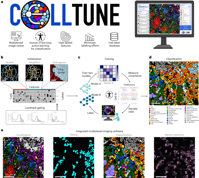
**Decoding the Cellular Microcosm: How Advanced Imaging is Revolutionizing Our Understanding of Tissues**
The microscopic world within our bodies has long been a frontier of immense complexity. For decades, pathologists relied on static snapshots and laborious manual analysis to interpret tissue biopsies. However, a technological renaissance in cellular imaging is dramatically changing this landscape, moving us from a state of observation to one of comprehensive, data-driven understanding. The latest research, as detailed in recent scientific literature, is enabling the simultaneous mapping of hundreds of cellular proteins, revealing the intricate choreography of cells within their native environments and unlocking new paradigms in diagnostics and research.
### The Rise of Hyper-Multiplexed Imaging
At the heart of this revolution is the development of **highly multiplexed imaging technologies**. These platforms have transcended the limitations of traditional methods, which were often restricted to detecting only a handful of markers at a time. Modern techniques are now capable of analyzing dozens, and in some cases over a hundred, distinct molecular signatures within a single tissue sample. This explosion in multiplexing capability is powered by a variety of innovative strategies:
* **Mass Cytometry (CyTOF):** This technology uses heavy metal tags instead of fluorescent dyes, allowing for the simultaneous measurement of numerous protein markers with minimal signal overlap and exceptional sensitivity.
* **Imaging Cyclic Staining (ICS/MICS):** By iteratively staining and stripping fluorescently labeled antibodies, these methods build up a complex multiplexed image one marker at a time, providing a powerful solution for deep phenotyping.
* **Spectral Imaging & AI-Driven Analysis:** Advanced algorithms, including machine learning and deep learning, are now essential for parsing the massive, complex datasets generated by these experiments. These tools can identify cell types, quantify protein expression, and map intricate cellular neighborhoods with unprecedented accuracy.
This technological leap is beautifully illustrated by platforms like **MIBI-TOF (Multiplex Ion Beam Imaging Time-of-Flight)**, which can profile dozens of elements simultaneously, and **CODEX** and **MACSima**, which offer high-resolution, multiplexed imaging of formalin-fixed tissues.
### From Pixels to Pathways: Understanding the Tumor Microenvironment
One of the most profound impacts of these advances is in the field of oncology. Tumors are not simply masses of malignant cells; they are complex ecosystems comprising immune cells, stromal cells, and a dynamic molecular landscape that dictates the tumor’s response to therapy and its potential for metastasis.
* **Revealing Immune Dynamics:** High-plex imaging has shown that the “tumor microenvironment” is a critical determinant of patient outcomes. For example, technologies like **IBEX** and **RAMCC** enable the deep phenotyping of immune cells within tumors, revealing a structured and organized immune landscape at the invasive front of cancers like colorectal and triple-negative breast cancer. This organization is a powerful predictor of patient prognosis.
* **Identifying Therapeutic Targets:** By mapping the precise location of immune checkpoints (like PD-1) and tumor-specific antigens, researchers can identify why some patients respond to immunotherapies while others do not. Studies using **MICS** and other platforms have identified specific combinatorial target pairs that are crucial for effective CAR T-cell treatment of solid tumors, offering a roadmap for next-generation therapies.
* **Understanding Cellular States:** Beyond just identifying cell types, these tools can reveal the functional state of cells. Research has linked specific, molecule-defined “neighborhoods” of interacting cells to disease states, such as the myeloid-inflamed tumor-immune complex associated with poor prognosis in certain cancers.
### The Power of Artificial Intelligence
The sheer volume of data generated by these multiplexed experiments would be impossible to analyze manually. This has led to an inseparable partnership between advanced imaging and artificial intelligence. Machine learning models are trained on vast image datasets to perform tasks such as:
* **Automated Cell Segmentation and Classification:** Algorithms like **Cellpose** and tools developed by projects like **cellxgene** can accurately identify and categorize thousands of cells in a complex image.
* **Phenotypic Analysis:** AI can discover subtle, data-driven phenotypic patterns in diseased cells (e.g., in Acute Myeloid Leukemia) that correlate with specific prognoses, revealing insights that would be invisible to the human eye.
* **Predictive Modeling:** These models can learn from the spatial relationships between different cell types and protein expression levels to predict clinical outcomes or response to treatment.
### Conclusion: A New Era of Precision Spatial Biology
The integration of cutting-edge imaging, multiplexing strategies, and artificial intelligence has propelled biological research into a new era of spatial resolution and molecular depth. We are no longer just looking at tissues; we are deconstructing them into their fundamental cellular and molecular components with a level of precision that was once the stuff of science fiction.
This field is rapidly evolving from a purely research-oriented toolset into a cornerstone of clinical diagnostics and drug development. As these technologies become more accessible and their analytical pipelines more refined, they promise to transform our understanding of human disease, leading to more personalized and effective treatments. The future of pathology is not just in looking at cells, but in understanding the entire complex narrative they tell together.
—
### Frequently Asked Questions (FAQ)
**Q1: What is the main advantage of multiplexed imaging over traditional methods?**
A1: The primary advantage is the ability to analyze dozens to hundreds of different molecular markers simultaneously within a single tissue sample. This provides a much more comprehensive and context-rich view of cellular identity and interactions than traditional methods, which are typically limited to a few markers at a time.
**Q2: How is artificial intelligence (AI) used in this field?**
A2: AI, particularly machine learning and deep learning, is essential for analyzing the massive, complex datasets generated by multiplexed imaging. It is used for automating cell segmentation, classifying cell types based on their protein expression and spatial location, identifying subtle phenotypic patterns, and building predictive models for disease outcomes and treatment response.
**Q3: What are some real-world applications of this technology in cancer research?**
A3: In cancer research, these technologies are used to:
* Map the tumor-immune microenvironment to understand why some tumors respond to immunotherapy and others don’t.
* Identify specific cellular “neighborhoods” and protein interactions associated with poor patient prognosis.
* Discover novel combinatorial targets for next-generation cellular therapies like CAR T-cells.
* Develop predictive diagnostics based on the spatial organization of immune cells within a tumor.
**Q4: What are some of the key technologies enabling this revolution?**
A4: Key technologies include mass cytometry (e.g., CyTOF), imaging cyclic staining (e.g., MICS, MACSima), and advanced spectral imaging platforms (e.g., IBEX). These are coupled with powerful data analysis tools and image analysis software.
**Q5: Is this technology being used in clinical settings yet?**
A5: While many applications are still in the research phase, the field is rapidly moving towards clinical translation. These technologies are increasingly being used for advanced diagnostics, particularly in oncology and immunopathology, to provide information that directly informs treatment strategy. The development of standardized analysis pipelines and the decreasing cost of the technology are accelerating its adoption in clinical laboratories.
