



# The Role of Artificial Intelligence in Modern Radiation Oncology: Transforming Cancer Treatment Through Data-Driven Innovation
## Introduction
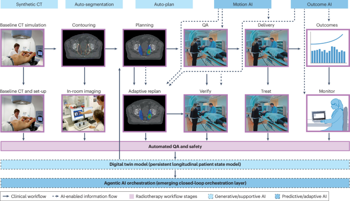
Radiation therapy remains one of the most widely used treatments for cancer, with approximately half of all cancer patients receiving some form of radiotherapy during their care. Over the decades, advances in imaging, linear accelerator technology, and treatment planning have steadily improved outcomes. However, the complexity of modern radiotherapy — from precise tumor targeting to minimizing damage to healthy tissues — has created growing demand for tools that can enhance speed, accuracy, and personalization. Artificial intelligence (AI) has emerged as a powerful ally in meeting this demand, offering solutions across virtually every stage of the radiation oncology workflow.
The integration of AI into radiation oncology represents a paradigm shift, moving from purely manual, experience-dependent processes toward data-driven, automated, and predictive approaches. This article explores how machine learning, deep learning, and emerging foundation models are reshaping treatment planning, quality assurance, outcome prediction, and clinical decision-making in radiation therapy.
—
## From Traditional Planning to Intelligent Treatment Design
Historically, radiation treatment planning was a labor-intensive process performed by dosimetrists and medical physicists. The goal was to design beams and dose distributions that would deliver a lethal dose to the tumor while sparing surrounding organs at risk. With the advent of intensity-modulated radiation therapy (IMRT) and volumetric modulated arc therapy (VMAT), the complexity of plan optimization increased dramatically, requiring sophisticated inverse planning algorithms and extensive manual adjustment.
AI has introduced the possibility of automating much of this workflow. Knowledge-based planning systems, which use historical plan data to suggest optimal dose distributions for new patients, have been in development since the late 1990s. Early neural network models demonstrated the ability to predict dose-volume histograms for organs at risk, laying the groundwork for fully automated treatment planning. More recently, deep reinforcement learning frameworks have been applied to treatment plan optimization, enabling systems to learn optimal beam configurations and dose patterns through iterative simulation.
These AI-driven approaches have shown particular promise in specific cancer sites. For prostate cancer, automated planning systems using hierarchical deep learning architectures can predict three-dimensional dose distributions directly from patient anatomy. In head and neck cancer, densely connected U-net architectures have been used to generate highly accurate dose predictions. For cervical cancer, deep reinforcement learning has been applied to brachytherapy treatment planning, demonstrating the ability to optimize dose distributions in a fraction of the time required for traditional manual approaches.
The clinical value of these systems lies not only in their speed but also in their consistency. By reducing inter-planner variability, AI-assisted planning can help ensure that every patient receives a treatment plan of comparable quality, regardless of which institution or planner is involved.
—
## AI-Assisted Contouring and Image Segmentation
One of the most time-consuming steps in radiotherapy preparation is the delineation of tumors and organs at risk on medical images, a process known as contouring or segmentation. Radiation oncologists must manually identify tumor boundaries and critical structures on CT and MRI scans, a task that is both subjective and variable between practitioners.
Deep learning-based auto-contouring systems have transformed this process. Convolutional neural networks trained on large datasets of annotated medical images can now segment organs at risk and lymph node regions with clinical-grade accuracy. These systems have been validated across multiple cancer types, including head and neck, lung, prostate, and brain tumors. Commercial auto-contouring tools are now widely available and have been shown to significantly reduce contouring time while maintaining or improving consistency.
However, the deployment of AI contouring tools is not without challenges. Studies have highlighted the importance of rigorous validation, noting that auto-contouring systems can exhibit population-specific biases if trained on datasets that do not represent the diversity of the patient population. Clinical acceptance studies have shown that while AI-generated contours often require only minor adjustments, they may not always match the nuanced judgment of an experienced radiation oncologist. Ongoing efforts focus on developing human-in-the-loop workflows where AI assistance speeds up the process while the clinician retains final oversight.
—
## Synthetic Imaging and Dose Calculation Advances
Magnetic resonance imaging (MRI) offers superior soft-tissue contrast compared to computed tomography (CT), making it increasingly attractive for radiation therapy guidance, particularly in prostate and brain cancers. However, dose calculation algorithms for photon and proton therapy require CT-derived electron density information, which MRI does not directly provide. This limitation has motivated the development of AI-based synthetic CT generation from MRI images.
Generative adversarial networks (GANs) and other deep learning architectures have been developed to create synthetic CT images from MRI scans with sufficient accuracy for dose calculation. Multiple studies have validated these approaches across different anatomical sites, including the pelvis, brain, and head and neck region. The emerging capability for synthetic-CT-free dose calculation in MRI-guided radiotherapy could simplify workflows, reduce scan times, and enable more precise adaptive treatment strategies.
These imaging advances are particularly relevant for MR-guided adaptive radiation therapy, where treatment plans are modified in real time based on daily imaging. By eliminating the need for co-registered CT scans, synthetic imaging approaches could make MRI-only workflows more practical and widely accessible.
—
## Predicting Treatment Outcomes with Machine Learning
Beyond the planning and delivery phases, AI is making significant inroads into outcome prediction — estimating how a patient will respond to radiotherapy based on their clinical, dosimetric, and imaging data. Machine learning models have been developed to predict a range of treatment outcomes, including tumor control, survival, and treatment-related toxicities such as radiation pneumonitis, xerostomia, and skin reactions.
Early work using multivariable modeling and statistical learning techniques demonstrated that dosimetric parameters (such as dose-volume metrics for organs at risk) and clinical factors (such as patient age and comorbidities) could be combined to predict treatment outcomes with meaningful accuracy. More recent studies have incorporated radiomics — the extraction of quantitative features from medical images — and even genomic data to build more comprehensive predictive models.
Deep learning architectures have further expanded these capabilities. Transformer-based models have been explored for survival prediction using patient-reported outcomes, while multiomics integration approaches have combined imaging, dosimetry, and molecular data in joint prediction frameworks. These models are particularly valuable in identifying patients at high risk of complications, enabling personalized treatment modifications or closer monitoring.
—
## Digital Twins and Personalized Simulation
The concept of a digital twin — a computational representation of a patient’s anatomy and physiology that can be used to simulate treatment responses — has gained significant attention in radiation oncology. Digital twins integrate patient-specific imaging, dosimetric, and biological data to create a virtual model that can be used to test different treatment strategies before they are applied in clinical practice.
In radiation therapy, digital twins can be used to simulate how a tumor and surrounding normal tissues will respond to different dose fractionation schemes, beam configurations, or systemic therapy combinations. Adaptive radiotherapy, where treatment plans are modified during the course of therapy based on anatomical changes, can be enhanced through digital twin simulations that predict optimal adjustments.
Research has demonstrated the feasibility of digital twin frameworks for adaptive treatment planning, particularly in proton therapy, where the sensitivity of dose deposition to anatomical changes makes real-time optimization especially valuable. Challenges remain, however, in terms of data integration, computational demands, and the need for rigorous clinical validation before these models can be deployed in routine practice.
—
## Generative AI and Large Language Models in Radiation Oncology
The rapid advancement of generative AI and large language models (LLMs) has opened new frontiers in radiation oncology. These models, which can understand and generate human language as well as process multimodal data, have potential applications ranging from clinical documentation to treatment planning assistance and patient education.
Large language models have been explored for automating toxicity extraction from oncology trial reports, summarizing complex treatment plans, and assisting with quality assurance documentation. In educational settings, LLMs have been evaluated as tools for helping patients understand their diagnosis and treatment options, as well as for training radiation oncology residents.
Generative AI has also been applied to the creation of synthetic data, which can help address the challenge of limited training datasets in rare cancer types or underrepresented populations. Foundation models — large, pre-trained AI systems that can be fine-tuned for specific tasks — are being investigated for their potential to serve as general-purpose tools across multiple radiation oncology applications, from image segmentation to outcome prediction.
However, these technologies come with important caveats. Concerns about hallucination, bias, data privacy, and the interpretability of model outputs must be carefully managed. The regulatory landscape for AI in medicine is evolving rapidly, with frameworks being developed by agencies such as the U.S. Food and Drug Administration (FDA) and the European Union to ensure that AI-enabled medical devices meet standards for safety, efficacy, and equity.
—
## Quality Assurance and Safety Considerations
As AI becomes more deeply integrated into radiation therapy workflows, robust quality assurance (QA) processes are essential. AI-based applications in radiation therapy — from auto-contouring to automated planning and outcome prediction — must be subject to the same rigor as any other clinical tool, and in many cases, even greater scrutiny.
Guidelines from professional societies have emphasized the importance of clinical validation, transparent reporting of model performance, and ongoing monitoring after deployment. Key considerations include ensuring that AI tools perform consistently across different patient populations, imaging equipment, and clinical scenarios. Fail-safe mechanisms, such as human oversight and override capabilities, remain critical components of any AI-assisted clinical workflow.
Researchers have developed deep learning-based QA systems that can predict treatment plan quality metrics or identify delivery errors from log file data. These tools have the potential to catch problems before they affect patients, adding an additional layer of safety to the treatment process.
—
## Ethical, Regulatory, and Educational Dimensions
The ethical implications of AI in radiation oncology are multifaceted. Issues of algorithmic bias, fairness, and equity must be addressed to ensure that AI tools benefit all patients, regardless of demographic characteristics or the population on which the models were trained. Data privacy, informed consent, and the potential for AI to displace or augment human roles in the clinical workforce are additional areas of active discussion.
Regulatory frameworks are still catching up with the pace of technological innovation. The FDA has issued guidance on predetermined change control plans for AI-enabled medical devices, and the European Union’s AI Act provides a broader regulatory framework for high-risk AI applications. In radiation oncology specifically, joint guidelines from professional societies have been developed to standardize the development, validation, and reporting of AI models.
Education and training are equally important. As AI tools become standard in radiotherapy practice, the next generation of radiation oncologists, medical physicists, and dosimetrists must be proficient in understanding, evaluating, and using these technologies. Efforts are underway to develop dedicated AI curricula, online learning platforms, and hands-on training programs in radiation oncology departments worldwide.
—
## The Path Forward
Artificial intelligence is no longer a futuristic concept in radiation oncology — it is a present reality that is already influencing clinical workflows, research, and education. The coming years will likely see continued integration of AI across all aspects of radiotherapy, driven by advances in foundation models, multimodal data integration, and real-time adaptive capabilities.
Key challenges that must be addressed include the need for large, diverse, and high-quality training datasets; the development of standardized evaluation metrics and benchmarking tools; the establishment of clear regulatory pathways; and the cultivation of trust among clinicians and patients. Interdisciplinary collaboration between radiation oncologists, medical physicists, computer scientists, ethicists, and regulatory experts will be essential to realizing the full potential of AI in this field.
—
## Frequently Asked Questions (FAQ)
**What types of AI are used in radiation therapy?**
Machine learning, deep learning, reinforcement learning, generative AI, and large language models are all used across different stages of the radiation therapy workflow, from image segmentation and treatment planning to outcome prediction and clinical documentation.
**Can AI replace radiation oncologists?**
No. AI is designed to assist and augment clinical decision-making, not replace human expertise. Radiation oncologists provide the clinical judgment, contextual understanding, and final decision authority that AI systems cannot replicate.
**How accurate are AI-based auto-contouring tools?**
Modern deep learning-based auto-contouring systems achieve accuracy levels that are comparable to expert human contouring for many anatomical structures. However, performance varies by cancer site, imaging modality, and the specific AI model used, and clinical review by a trained specialist remains essential.
**What is synthetic CT, and why does it matter?**
Synthetic CT is an AI-generated CT-like image created from an MRI scan. It matters because MRI offers superior soft-tissue visualization, and synthetic CT can enable MRI-only radiotherapy workflows, reducing the need for multiple scans and streamlining treatment planning.
**Are there regulatory standards for AI in radiation therapy?**
Yes. Regulatory bodies such as the FDA and the European Union have established frameworks for AI-enabled medical devices. Professional societies have also published guidelines for the development, validation, and clinical reporting of AI models in radiation therapy.
**What are the biggest challenges for AI adoption in radiation oncology?**
Key challenges include ensuring data diversity and reducing algorithmic bias, obtaining rigorous clinical validation, maintaining patient data privacy, integrating AI tools into existing clinical workflows, and training the next generation of clinicians to work effectively with these technologies.
**What is a digital twin in the context of radiation therapy?**
A digital twin is a computational model of a patient’s anatomy and disease that can be used to simulate treatment outcomes under different scenarios. In radiation therapy, digital twins can help optimize treatment plans and guide adaptive strategies in real time.
—
## Conclusion
Artificial intelligence is fundamentally reshaping the landscape of radiation oncology. From intelligent treatment planning and automated contouring to predictive outcome modeling and digital twin simulations, AI is enhancing every phase of the radiotherapy workflow. As these technologies mature and become more widely available, they hold the promise of making radiation therapy more precise, efficient, equitable, and personalized than ever before. Realizing this potential will require continued investment in research, thoughtful regulation, robust quality assurance, and a commitment to educating both current and future clinicians. The journey is well underway, and the integration of AI into radiation therapy represents one of the most exciting developments in modern cancer care.
Thank you for reading
