



**Harnessing Non-Destructive 3D Pathology to Revolutionize Diagnostic Precision**
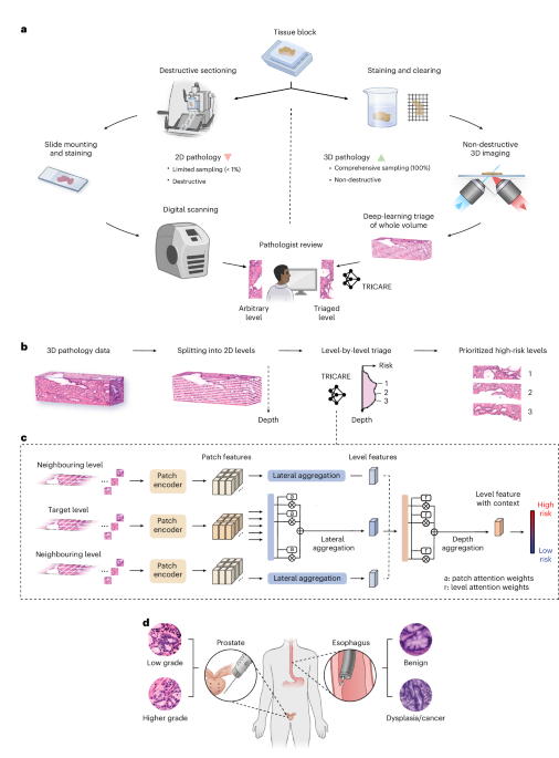
The landscape of pathological diagnostics is undergoing a profound transformation, driven by the convergence of advanced imaging technologies and artificial intelligence. Traditional histopathology, while the gold standard for disease diagnosis, is inherently limited by its reliance on two-dimensional (2D) sections of tissue. This approach requires pathologists to mentally reconstruct complex 3D structures from thin slices, a process that is time-consuming, subject to inter-observer variability, and can lead to diagnostic inaccuracies. The emergence of non-destructive 3D pathology offers a paradigm shift, enabling the comprehensive analysis of intact specimens in three dimensions, thereby providing a more holistic and accurate view of disease.
**The Foundations of 3D Pathology**
The core principle of non-destructive 3D pathology is to visualize tissues in their native, volumetric state without the need for physical sectioning. This is achieved through a suite of advanced optical and imaging techniques. Key among these is **light-sheet microscopy**, particularly the open-top variant, which uses a thin sheet of light to illuminate a specimen plane-by-plane, minimizing photo-damage and enabling high-speed, high-resolution imaging of large tissues. This technology has been further refined through innovations such as multi-immersion objectives and hybrid microscope designs, which enhance resolution, field-of-view, and imaging depth.
Crucially, the integration of **tissue clearing** methods has been a game-changer. These protocols render tissues transparent by removing lipids and scattering molecules, allowing light to penetrate deeply. This enables the generation of detailed 3D maps of entire organs or even whole bodies, revealing the spatial architecture of cells and tissues with unprecedented clarity. When combined with advanced labeling techniques like fluorescent antibodies or genetic markers, these methods provide specific molecular and cellular information within this 3D context.
**The Role of Artificial Intelligence**
The immense data generated by 3D imaging presents a challenge that artificial intelligence (AI) is uniquely equipped to address. Machine learning and deep learning algorithms are being developed to analyze these complex datasets, augmenting the capabilities of pathologists. AI applications range from **automated triage**, where algorithms prioritize suspicious regions within a 3D volume for closer examination, to **semantic segmentation**, where models can precisely identify and delineate specific cell types, such as cancerous glands in a prostate biopsy or tumor boundaries in a lymph node.
These AI tools are designed to reduce pathologist workload, minimize diagnostic errors, and uncover subtle patterns invisible to the human eye. For instance, research has demonstrated that AI can analyze nuclear features in 3D-prostate cancer specimens to predict disease aggressiveness, or detect micro-anatomical structures in cleared lungs to assess fibrosis. By acting as a “second pair of eyes,” AI enhances both the accuracy and efficiency of pathological workflows.
**Clinical and Research Impact**
The implications of this technological shift are far-reaching. In oncology, 3D pathology allows for more precise tumor margin assessment during breast-conserving surgery, potentially reducing the need for repeat procedures. It offers a comprehensive view of tumor heterogeneity and its relationship with the surrounding microenvironment, which is critical for understanding disease progression and treatment response. Furthermore, in complex organs like the prostate, 3D analysis provides a more accurate representation of cancer foci compared to traditional 2D sampling, leading to better risk stratification and more informed treatment decisions.
Research is also expanding the frontiers of biological discovery. 3D imaging atlases, such as those created for Barrett’s esophagus or colorectal cancer, are providing new insights into the spatial organization of disease. This spatial information is crucial for understanding cell-cell interactions, immune responses, and the evolution of pre-cancerous lesions, ultimately paving the way for more personalized and effective therapies.
—
### **Frequently Asked Questions (FAQ)**
**Q1: What is non-destructive 3D pathology?**
A1: Non-destructive 3D pathology refers to a set of imaging techniques that allow for the visualization of entire biological specimens in three dimensions without physically cutting or sectioning them. Methods like light-sheet microscopy combined with tissue clearing enable pathologists and researchers to examine the full spatial architecture of tissues, preserving them for future analysis and providing a more complete diagnostic picture than traditional 2D histology.
**Q2: How does artificial intelligence (AI) contribute to 3D pathology?**
A2: AI algorithms are essential for managing and interpreting the vast and complex datasets generated by 3D imaging. They can automatically detect, segment, and quantify cellular and structural features within these volumes. This assists pathologists by triaging cases, highlighting areas of concern, and providing quantitative, objective measurements that improve diagnostic accuracy and workflow efficiency.
**Q3: What are the main benefits of 3D pathology over traditional methods?**
A3: The primary benefits include a more comprehensive assessment of tissue architecture, reduced inter-observer variability, and the ability to detect abnormalities that may be missed in 2D slices. It provides a spatially accurate context for diagnosis, leading to better understanding of disease and more informed clinical decisions. It also preserves the specimen for further molecular or genetic testing.
**Q4: What are some common tissue clearing techniques used in 3D pathology?**
A4: Common clearing methods include CUBIC, SeeDB, and SHANDI, which use various chemical solutions to remove lipids and water from tissues, making them transparent and optically accessible. These are often tailored for specific tissue types, such as the “See-Deep” method for clearing deep tissues or refractive index matching techniques for optimal light penetration in microscopy.
**Q5: Is 3D pathology currently being used in clinical diagnostics?**
A5: While the technology is still evolving and being validated, non-destructive 3D pathology is increasingly being integrated into clinical workflows, particularly in specialized centers. Its adoption is growing rapidly, driven by advancements in hardware, software, and AI, with applications in cancer diagnostics, surgical pathology, and research.
—
### **Conclusion**
Non-destructive 3D pathology represents a monumental leap forward in our ability to visualize and understand disease at a systems level. By overcoming the fundamental limitations of 2D histology, it provides an unparalleled view of tissue architecture and pathology. The synergistic integration of this advanced imaging with artificial intelligence is not merely an incremental improvement but a revolutionary step toward precision medicine. As these technologies continue to mature and become more accessible, they hold the promise of transforming diagnostics, improving patient outcomes, and unlocking new frontiers in biomedical research. The future of pathology is not just in looking at slides, but in exploring the intricate, three-dimensional world of human health and disease.
